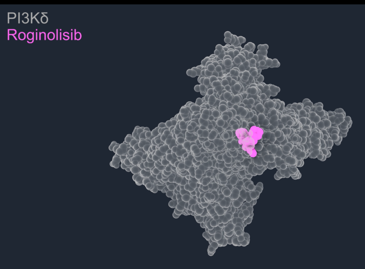
A new publication in Nature Communications reveals how roginolisib uniquely locks the cancer-driving enzyme PI3Kδ in an inactive state, differentiating the drug from other PI3Kδ inhibitors.
PI3Kδ has long been recognized as an important therapeutic target in cancer, but previous generations of inhibitors have been severely limited by tolerability issues, associated in part with the way they bind to the enzyme. In this mechanistic study, researchers from the University of Geneva and the Max Planck Institute of Biophysics in Frankfurt used X-ray crystallography, molecular dynamics simulations and hydrogen-deuterium exchange mass spectrometry to investigate how roginolisib interacts with PI3Kδ. They revealed a novel binding mode: roginolisib locks the enzyme into an inactive conformation by uniquely stabilizing its catalytic C-terminal helix kα12.
We spoke with Lars van der Veen, co-author of the study and Chief Scientific Officer of iOnctura, about what the findings mean and why they could matter for the future of PI3Kδ inhibition.
Q.
What is the key finding of this paper?
The central finding is that roginolisib appears to inhibit PI3Kδ in a unique manner, that is very different from previously approved PI3Kδ inhibitors.
Most PI3K inhibitors have been developed to interact with the ATP-binding pocket of the enzyme. What we found is that roginolisib not only binds within this pocket but also appears to induce and stabilize an inactive conformation of PI3Kδ involving the enzyme’s C-terminal regulatory helix, known as kα12.
To our knowledge, stabilization of this helix as a mechanism of PI3Kδ inhibition has not previously been described.
Q.
Why is that important?
Kinases such as PI3K are dynamic proteins. They constantly move between active and inactive conformations. Many inhibitors bind to the active state of the protein. For example, this appears to be the case with idelalisib, a PI3Kδ inhibitor approved for CLL.
Our data suggests that roginolisib is different. It preferentially targets and stabilizes the inactive form of PI3Kδ. In simple terms, rather than trying to block the enzyme once it is active, it may help keep the enzyme switched off. That concept is interesting because inactive conformations often contain structural features that differ between related enzymes, for example the different isoforms of PI3K. Those differences can potentially be exploited to improve selectivity and improve upon first-generation PI3Kδ drugs such as idelalisib.
Q.
What are the major differences between roginolisib and first-generation inhibitors?
First-generation PI3Kδ inhibitors demonstrated the therapeutic potential of targeting this pathway, but their clinical use was often limited by toxicities including colitis, infections and liver injury. These therapies broadly suppressed PI3Kδ signaling, making it difficult to separate the disease-driving activity of the pathway from the normal signaling required for healthy immune function.
In cancers such as CLL, PI3Kδ signaling is persistently activated by the tumor’s own survival mechanisms. Healthy immune cells, however, rely on the same pathway to perform their normal functions. Our goal with roginolisib is to inhibit the pathogenic signaling that promotes cancer whilst preserving the signaling associated with healthy immune function. The emerging clinical profile of roginolisib is encouraging. Unlike idelalisib, roginolisib demonstrates a highly selective kinase profile. Whether the conformation-selective mechanism described in this study contributes to that differentiation remains an active area of investigation.
Q.
How does the binding mechanism differ between roginolisib and idelalisib?
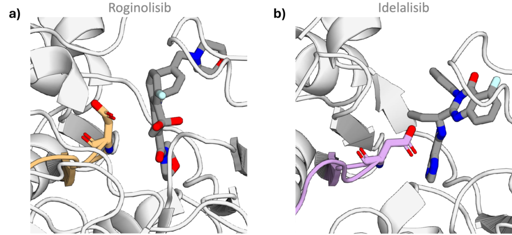
The structural studies showed that the two molecules interact with PI3Kδ in distinct ways. Idelalisib preferentially binds the active form and thereby competes with ATP, the energy source of the enzyme, for binding. Conversely, roginolisib stabilizes the kα12 helix in a folded, inactive arrangement over the catalytic site. Because ATP cannot bind to the inactive form of the enzyme, the binding of roginolisib is not competitive with ATP.
This is important as it means roginolisib better retains its potency in cells: A lower concentration of drug can be used to achieve the desired effect.
Q.
Can you explain kα12 in simple language?
A useful way to think about PI3Kδ is as an enzyme with a built-in regulatory gate. That gate is formed by a structural element called the kα12 helix, located at the C terminal end of the protein. To activate PI3Kδ, kα12 hinges away from the active site and helps position the enzyme at the cell membrane, where it can convert its substrate PIP2 into PIP3, a key signaling molecule that drives PI3K pathway activity. Our study suggests that roginolisib effectively locks this gate.
Roginolisib promotes interactions between the acidic residues Asp897 and Asp911within the active site and the basic residues Lys1040 and Arg1043 of the kα12 helix. Molecular dynamics simulations suggest that this network of interactions stabilizes kα12 in the folded, “locked” inactive conformation. Interestingly, Lys1040 and Arg1043 are also the residues that appear to help the active enzyme engage its lipid substrate during normal signaling. So, in the presence of roginolisib, these residues aren’t available to partake in active signaling.
In contrast, the same interaction network was not observed with idelalisib. Our simulations suggested that kα12 remains more open and flexible in the presence of idelalisib, consistent with the inhibition of the active kinase.
Q.
Why might this lead to better selectivity?
One of the most intriguing observations is that several of key residues involved in roginolisib’s interaction with kα12 are not conserved across all PI3K family members. They are found in the δ and β isoforms but not in α or γ.
Conversely, historical PI3Kδ inhibitors only engage residues that are highly conserved in the ATP-binding pocket. This makes them inherently less selective for the δ isoform.
Q.
The research shows differential activity of roginolisib and idelalisib on PIP3. What does this mean and why does it matter?
When PI3Kδ is activated, it converts PIP2 into PIP3, which then triggers signaling events that promote cell survival, proliferation and immune-cell function. If a PI3Kδ inhibitor is working effectively, one of the most direct ways to demonstrate that is to show reduced PIP3 production.
This research moved beyond structural biology and measured PIP3 directly in primary CLL patient samples. The research showed that roginolisib produced stronger and more consistent suppression of PIP3 than idelalisib under the conditions tested.
Q.
Does this tell us anything about biology or clinical activity?
This is where it is important not to over-interpret the data. The paper does not prove that the kα12 mechanism causes any specific clinical benefit.
It does not establish a direct causal link between the structural observations and patient outcomes.
What it does provide is a mechanistic framework that may help explain several previously observed characteristics of roginolisib, including its high isoform selectivity, its ability to maintain activity under high ATP concentrations, and the robust suppression of PIP3 observed in patient-derived samples.
We propose that stabilizing the inactive form of PI3Kδ could represent a fundamentally different way of modulating the pathway. Whether that ultimately translates into distinct biological or clinical effects remains an area for ongoing investigation.
Q.
What is the broader significance of the work?
That question matters because PI3Kδ has already proven to be an important therapeutic target. First-generation inhibitors such as idelalisib demonstrated that blocking this pathway can benefit patients with diseases such as CLL, but their use was ultimately limited by toxicities including colitis, infections and liver injury. For many patients, these side effects made it difficult to remain on treatment long enough to achieve the full benefit of therapy.
The mechanism described in our study offers a different way of targeting PI3Kδ. By engaging the enzyme through a distinct binding mode associated with the inactive form of the enzyme, it establishes a new framework for selectively inhibiting the pathway while potentially avoiding some of the limitations associated with earlier generations of inhibitors.
The key question, of course, is whether this scientific insight can translate into better outcomes for patients. That is now being tested in the clinic. Roginolisib has been administered to more than 200 patients across multiple clinical studies in both solid tumors and hematological malignancies, as a monotherapy and in combination with targeted therapies including JAK and BCL-2 inhibitors. Through these studies, we hope to demonstrate that a differentiated approach to PI3Kδ inhibition can enable patients to benefit from prolonged pathway suppression with an improved tolerability profile compared with earlier generations of inhibitors.
Source